HLA-A*02:01 & B2M complex
Description
Human leukocyte antigen A (HLA-A) and beta 2 microglobulin (B2M) are components of the class I major histocompatibility complex (MHC-I), involved in presenting peptide antigens to the immune system. They primarily display viral and tumor-derived peptides on antigen-presenting cells for recognition by alpha-beta T cell receptors (TCRs) on HLA-A-restricted CD8+ T cells. This process guides antigen-specific T cell immune response aimed at eliminating infected or transformed cells. HLA-A may also present self-peptides originated from the signal sequence of secreted or membrane proteins, although T cells specific for these peptides are usually inactive to prevent autoreactivity. Both the peptide and the MHC molecule are recognized by TCR, the peptide determines fine specificity of antigen recognition and MHC residues account for MHC restriction in T cells. Typically HLA-A presents intracellular peptide antigens of 8 to 13 amino acids that arise from cytosolic proteolysis via IFNG-induced immunoproteasome or endopeptidase IDE/insulin-degrading enzyme. It can bind various peptides containing allele-specific binding motifs, which are mainly defined by anchor residues at position 2 and 9. Allele A*02:01 is a prevalent allele in human populations, that presents immunodominant viral epitopes derived from IAV M/matrix protein 1 (GILGFVFTL), HIV-1 env (TLTSCNTSV), HIV-1 gag-pol (ILKEPVHGV), HTLV-1 Tax (LLFGYPVYV), HBV C/core antigen (FLPSDFFPS), HCMV UL83/pp65 (NLVPMVATV), as well as tumor peptide antigens including MAGEA4 (GVYDGREHTV), WT1 (RMFPNAPYL) and CTAG1A/NY-ESO-1 (SLLMWITQC), all having hydrophobic amino acids at position 2 and C-terminal anchors in common. Exogenously applied M.tuberculosis EsxA or EsxA-EsxB binds B2M and decreases its export to the cell surface (total protein levels do not change), probably leading to defects in class I antigen presentation.
Product name | HLA-A*02:01 & B2M complex |
Species | Homo sapiens |
Expression system | HEK293 |
Buffer | PBS, pH 7.4 |
Delivery condition | Dry ice (-80°C) |
Delivery Time | 1 week if in stock; 4 weeks if production needed |
Storage condition | Store at -80°C |
Brand | BioMetas |
Applications | Immune System, antigen processing and presentation |
Aliases/Synonyms | peptide ready MHC, peptide free MHC, peptide receptive MHC |
Reference | |
Note | For research use only. Not suitable for clinical or therapeutic use. |
Size | 1mg, 5mg, 10mg, 50mg, 100mg |
Brand | BioMetas |
Product type | Antigen |
Expression system | HEK293 |
Applications | Immune System, antigen processing and presentation |
Contact Us for a Quote!
Data Gallery
Fig. 1.) 4-20% SDS-PAGE analysis
Recombinant protein was visualized by Coomassie Brilliant Blue R250 staining.
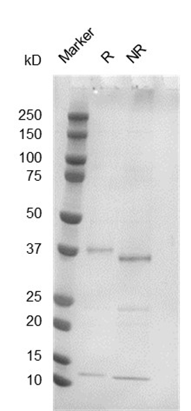
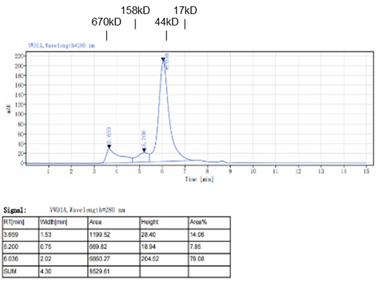
Fig. 2.) SEC-HPLC analysis
Column: Superdex 200 Increase 5/150 GL
Running buffer: 2xPBS, pH 7.4